

Representative of the IBS Director for the Laboratory
dr hab. Bożena Pawlikowska-Pawlęga, prof. UMCS
Department of Functional Anatomy and Cytobiology
Akademicka 19, 20-033 Lublin
room 133B, tel. 81 537 59 91
mail: bozena.pawlikowska-pawlega@mail.umcs.pl
Staff members
mgr Jarosław Pawelec, room 0101A, tel. 81 5375916; 660 342 397
mgr Barbara Zarzyka, room 33B, tel. 81 537 50 41.
History of the Institute Microscopy Laboratory
The Laboratory was initiated in 2001 at the Department of Comparative Anatomy and Anthropology as a Faculty unit subordinate to the Dean. In 2001-2012, it was headed by prof. dr hab. Antoni Gawron. On December 2019, dr hab. Bożena Pawlikowska-Pawlęga, who was in charge of the Electron Microscopy Laboratory until the end of September 2019, was appointed Representative of the Director of the Institute of Biological Sciences for the Institute Microscopy Laboratory.
General information
The laboratory participates in the scientific research of the Faculty of Biology and Biochemistry through work in the field of transmission and scanning electron microscopy. The unit also plays a didactic role in classes on the issues of microscopic techniques. This function is also complemented by annual participation in popularisation and promotion events of the Faculty for schools, such as the Lublin Festival of Science or the Open Door of the Faculty and in individual shows organised for schools.
The Laboratory is equipped with the following devices:
The Institute of Biological Sciences is also equipped with
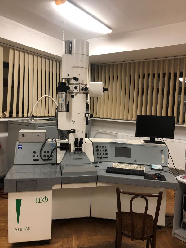
Transmission electron microscope (TEM) Zeiss LEO 912AB class 120kV [at present - out of service for technical reasons]
Premises: Laboratory of Electron Microscopy, room 0101A
Person in charge: mgr Jarosław Pawelec, room 125B, tel. 660 342 397
Auxiliary equipment: [ASC1]
TEM preparatory procedures:
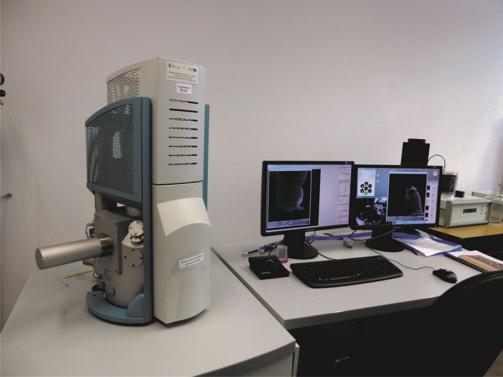
Tescan VEGA3 LMU scanning electron microscope (SEM) equipped with the variable vacuum mode and an optional Peltier freezing stage (up to -500C) for work with non-dehydrated organic samples
Premises: Department of Zoology and Nature Protection, room 152B
Person in charge: mgr Jarosław Pawelec, room 125B, tel. 660 342 397
Parameters:[ASC2]
Auxiliary equipment:
SEM preparatory procedures:
SEM examinations:
Motorised microscope stand
Premises: Institute Microscopy Laboratory, room 152B
Person in charge: mgr Jarosław Pawelec, room 125B, tel. 660 342 397
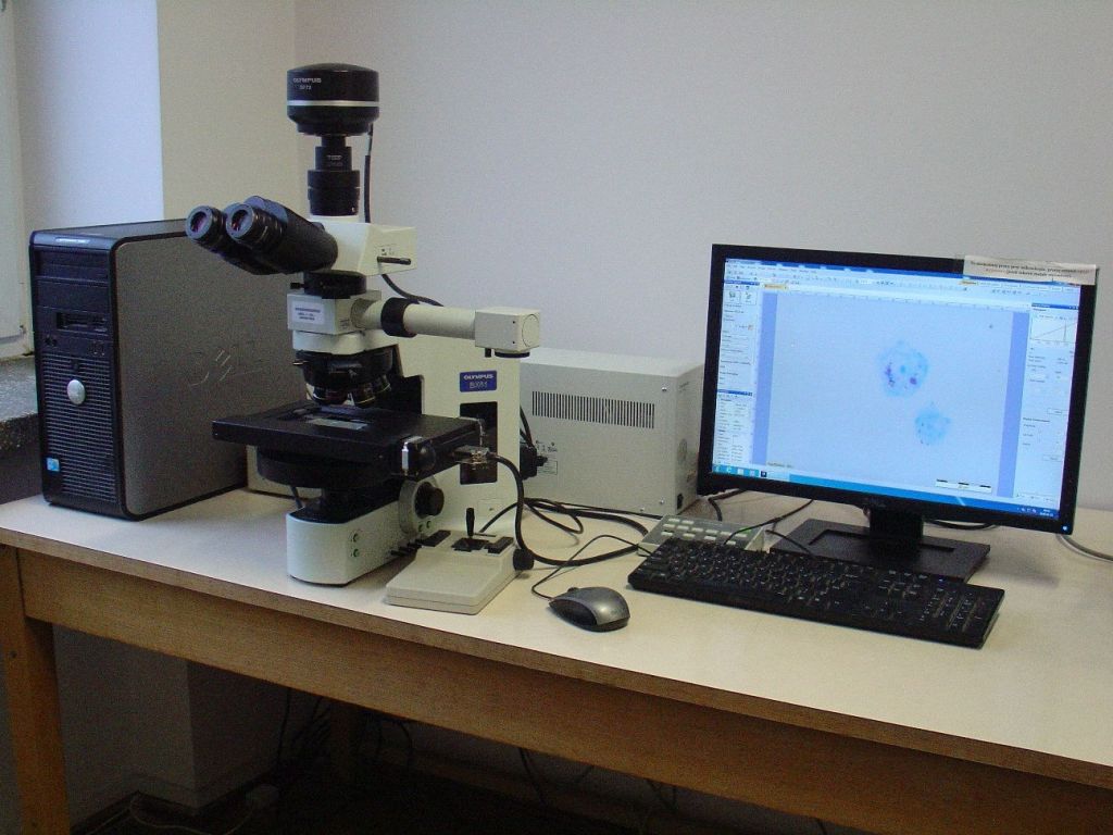
Olympus BX-61 biological microscope (with a camera and software)
Parameters: [ASC3]
Microscopic examinations:
Olympus SZX-16 stereo microscope (with a camera and software)
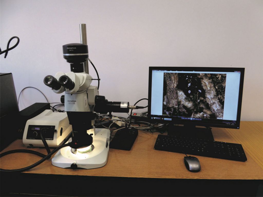
Parameters:[ASC4]
Microscopic examinations:
Zeiss Axiovert 200M confocal laser microscope with an LSM 5 Pascal scanning head
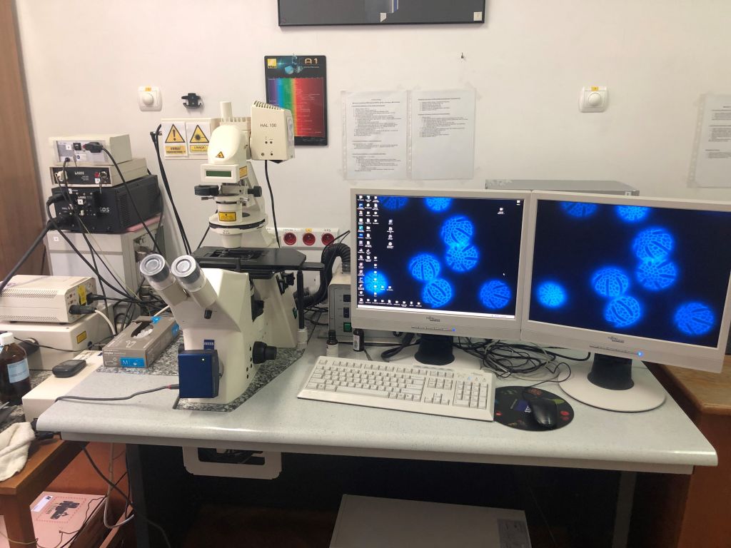
Premises: Department of Functional Anatomy and Cytobiology, room 126B
Person in charge: mgr Jerzy Wydrych, room 126B, tel. 81 537 59 98
Parameters and imaging techniques: [ASC5]
Services provided with the use of the device: biological, biophysical, biomaterial, and material examinations:
LSM 780 Zeiss laser confocal scanning microscope
Premises: Department of Molecular Biology, room 0106A
Person in charge: prof. Marek Tchórzewski, room 11A, tel. 81 5375956
dr Kamil Deryło, pokój 2A, tel. 81 5375922

The Environmental Laboratory of Intravital Cell Imaging is equipped with the LSM780 Zeiss laser confocal scanning microscopy system based on an Axio-Observer Z.1 microscope with two PMT detectors and a 32-channel GaAsP spectral detector. It has a class 3B laser system with 405, 458, 488, 514, 543, and 633 nm wavelengths. The microscope is equipped with dry lenses: EC Plan-Neofluar 10x/0.3 M27, Plan-Apochromat 20x/0.8 M27, and LD Plan-Neofluar 40x/0.6 Korr M27 as well as immersion lenses: Plan-Apochromat 40x/1.4 Oil DIC M27, LCI Plan-Neofluar 63x/1.3 Imm Korr DIC M27, and Plan-Apochromat 63x/1.4 Oil DIC M27. It also has a thermostatted Plexiglas chamber (controlled conditions - temperature, humidity, and CO2) providing cell lines with an appropriate environment for growth. The system is controlled with Zen2010 software. LSM780 is synchronised with the time-resolved fluorescence spectroscopy PicoQuant system consisting of a TCSPC (Time-Correlated Single-Photon Counting) module - PicoHarp 300, a two-channel SPAD (Single-Photon Avalanche Diodes) detector, and picosecond pulsed diode lasers with 440 and 485 nm wavelengths. Data acquisition and analysis are carried out using SymphoTime (PicoQuant) software. The laboratory is also equipped with an Axio Observer.D1 microscope with an AxioCam MRm Rev. 3Fire Wire digital camera and a 458-, 488-, and 514-nm laser system profiled for TIRF observations. The system is controlled with AxioVision Rel. 4.8.2 software.
The laboratory provides precise imaging of the structure of a living cell and tracking intracellular biological processes in a three-dimensional format, additionally implementing the time element (4D imaging). The system facilitates subcellular location of proteins and other molecules in static and dynamic systems. This research system is mainly intended for analysis of protein-protein, protein-ligand, and protein-nucleic acid interactions inside the living cell, on its surface, and in an in vitro system. The methods implemented in the system include FLIM (Fluorescence Lifetime Imaging Microscopy) imaging, spectral analysis with the use of an ultra-sensitive GaAsP detector, FRAP (Fluorescence Recovery After Photobleaching), FLIP (Fluorescence Loss In Photobleaching), FRET (Förster Resonance Energy Transfer), RICS (Raster-scanning Image Correlation Spectroscopy), FCS (Fluorescence Correlation Spectroscopy), anisotropy, and TIRF/FRET (Total Internal Reflection Fluorescence/Förster Resonance Energy Transfer).
Langmuir-Blodgett system coupled with a Brewster angle microscope and computer
Premises: Department of Cell Biology, Laboratory of Biospectroscopy, room 0118B
Person in charge: prof. Mariusz Gagoś, room 43B, tel. 81 5375904
Brewster's Angle Microscopy is one of the basic non-invasive techniques used for investigations of monolayers formed by water-insoluble amphiphilic substances on the water sub-phase surface. This technique is based on changes in the refractive index at the water (aqueous solution)/air interface induced by formation of a Langmuir monolayer.
Brewster angle microscopy (BAM) facilitates:
Equipment: a double-barrier Langmuir[ASC6] KSV trough (Helsinki, Finland) model 2000 with a total area of 700 cm2. A platinum Wilhelmy plate is used as a surface pressure sensor. Brewster angle microscope: ultraBAM, Accurion (Göttingen, Germany). The device uses a 50-mW laser emitting light with parallel polarisation (p) and a wavelength of 658 nm, an analyser, and a CCD camera. The module facilitates image detection with a resolution of 2 µm and 10x magnification. Radiation incident on the water/air interface is emitted by the Brewster microscope at an angle of 53.2°.
Ultrabam is a Brewster angle microscopy designed for the air/liquid interface. It provides direct visualisation of Langmuir monolayers or adsorbed layers. It also works with dielectric substrates such as glass, quartz, or similar materials. The nanofilm_ultrabam combines high resolution with focused real-time imaging. Advanced imaging optics provides fully focused images at 20-35 frames per second. The high performance camera and the specific calibration algorithm facilitate quantitative measurements of reflectivity. The adsorption kinetics or thickness alterations can thus be monitored. The device has a motorised analyser for visualisation of optical anisotropy caused by the long-range molecular orientation in monolayers. The powerful software makes operation easy and convenient. As a complete solution, the system consists of a computer, electronics, and all software required for initiation of measurements. The nanofilm_ultrabam is designed as a Brewster Angle Microscope that cannot be upgraded to an ellipsometer.
The Brewster Angle Microscopy (BAM) technique is successfully used for morphological and topological observation of monolayers. It provides information on the state of the monolayer, aggregation and formation of domains, and collapse mechanisms. It facilitates observation of phase separation and three-dimensional structures as well as detection of phase transitions. Before the analysis, a black glass plate is placed at the bottom of the Langmuir trough, which prevents radiation scattering through its Teflon bottom. After application of the monolayer into the surface of the pure subphase, the microscope is calibrated, which involves setting the Brewster angle as well as the image focus and contrast.
FTIR Vertex 70 spectrometer with a Hyperion 3000 infrared microscope
Premises: Department of Cell Biology, Laboratory of Biospectroscopy, room 0118B
Person in charge: prof. Mariusz Gagoś, room 43B, tel. 81 5375904
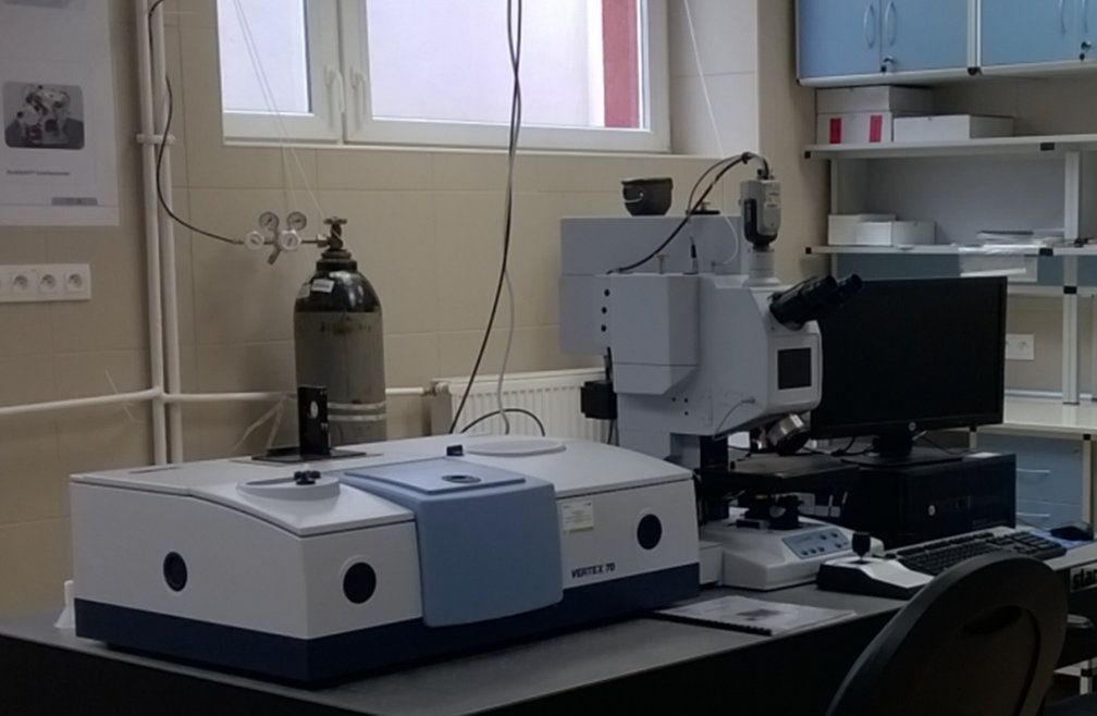
Analysis based on spectroscopic and micro-spectroscopic FT-IR measurements:
Basic parameters and measurement potential of the VERTEX 70 spectrometer:[ASC7]
1. Two detectors with automated selection and switching:
2. 60° Michelson interferometer:
3. Microscope coupled with the FT-IR spectrometer:
Measurement modes: reflection, transmission, and simultaneous measurements and observations in the visible mode, ATR;
Lenses: IR 15x for reflection and transmission measurements, IR 36x for reflection and transmission measurements, ATR 20x (ATR sealed lens with a 5-degree pressure from 0.8 to 8 N and electronic and visual control of the correct position of the crystal)
Detectors:
Thermostatted measurement bench with a microscope:
Additional equipment for the VERTEX 70 spectrometer:
1. Multifunctional attachment:
Measurement modes: ATR, external reflection, diffuse reflection;
Variable angle-of-attack range of at least 5-85;
ATR system with a ZnSe hemispherical crystal;
ATR system with a Ge hemispherical crystal;
Cell for measurements of liquid samples;
Thermostatted flow cell for measurement of liquid samples, thermostatic range at least 150°C;
Controller of the temperature of ATR attachments; temperature range at least 150°C;
Polariser;
Software for modelling parameters and digital simulation of spectra
2. Precision polariser:
Manually adjustable with a resolution of 1°;
Spectral range of work: from 5000 to 285 cm-1;
Transmission diameter: at least 22 mm
3. Flow measurement cell:
Whole windows made of ZnSe without apertures for sample insertion;
Layer thickness range - one spacer, at least 6-950 µm;
Operating temperature range from -80 to 200°C;
Possibility of thermostatting with an electric controller in a range up to at least 200°C;
Sample inlet - Luer type
4. Multi-angle horizontal ATR attachment ATR:
Measurement modes – ATR;
Variable angle-of-attack in the range of at least 20-70° adjustable at 1 ° increments;
Depth of sample penetration: from 0.5 to 10 µm;
Number of reflections: from 3 to 12;
45° ZnSe crystals and 45° Ge crystals in internal and external reflection modules;
Thermostatted flow cell module with a controller operating in the range of at least up to 120°C
5. Multi-reflection ATR attachment
Measurement modes – ATR;
Number of reflections: at least 10;
45°ZnSe crystal in an external reflection measurement module;
45° ZnSe thermostatted crystal in an internal reflection measurement module;
Possibility of measurement of liquid, solid, powder, volatile-solvent samples and samples with large sizes that do not fit in the measurement chamber
Leica DM 4000B phase-contrast and fluorescence microscope with LAS V3.1 acquisition, interactive measurement, and image analysis module
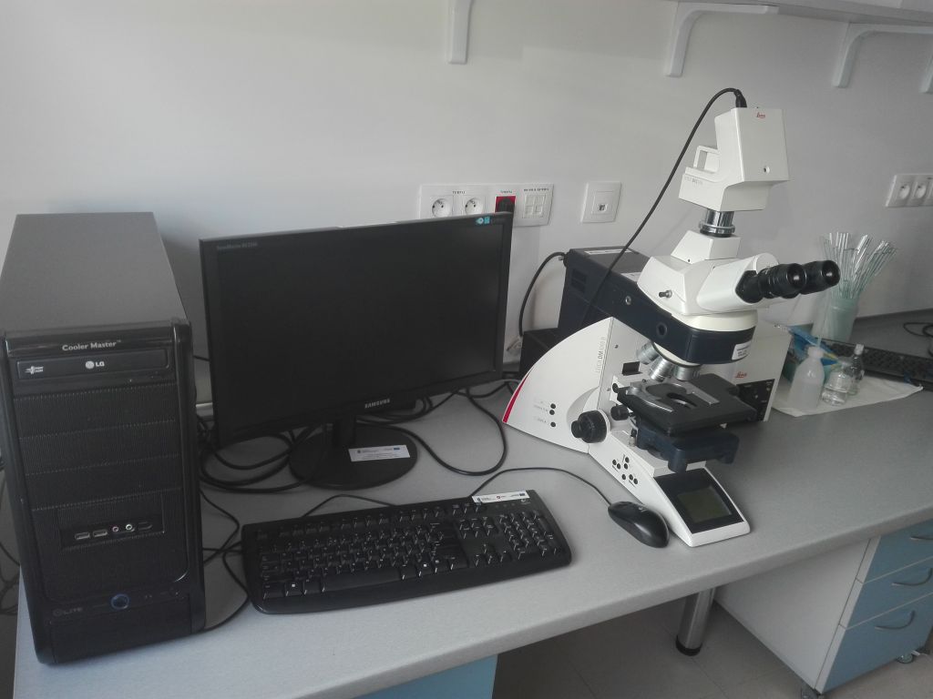
Premises: Department of Cell Biology, room 31B
Person in charge: prof. Mariusz Gagoś, room 43B, tel. 81 5375904
Analyses carried out with the use of the Leica DM 4000B phase-contrast and fluorescence microscope:
Basic parameters of the Leica DM 4000B phase-contrast and fluorescence microscope [ASC8]
Lenses (Plan Semi-apochromat Fluotar class):
Light source - a 100W halogen lamp
Control - function of automatic correction of illumination settings after changes in the magnification introduced by the user; manual correction of lighting and automatic saving of the change; motorised spherical and rectangular field aperture (for work with the camera); automatic correction of field aperture settings after changes in the magnification introduced by the user, manual aperture correction and automatic saving of the change; motorised aperture; automatic aperture correction function after changes in the magnification introduced by the user; possibility of manual correction of the aperture and automatic saving of the change
Illumination module: external light source based on a min. 150W metal-halide lamp with a 5-position motorised filter changer Leica EL6000
Fluorescence filters, broadband classes A, D, I3, N2.1
Digital camera: a Peltier actively cooled camera; aluminium housing with a fan; maximum resolution 12 Megapixel (4080 x 3072); min. 2/3 inch CCD matrix; pixel size min. 6.45 x 6.45 µm; active area min. 8.8 x 6.6 mm; exposure time min. from 0.25 ms. to 600 s; S/N ratio 2000:1 (66 dB); a 14-bit AD converter; 42-bit colour depth; 0.70 x C-mount connector; FireWire interface for connection with the Leica DFC500 computer
External PC computer with the Windows operating system; quad-core processor, clock-rating at least 2GHz, 4GB RAM, HDD 500GB, DVD R/RW DL; graphics card compatible with the camera control program at least 512MB, processor clock-rating at least 600MHz; PCI-Express 16x connector; USB 6x; power supply minimum 600W; minimum performance 80%, Fire Wire, Ethernet 1Gbps, Windows XP Pro, or Windows 7 Pro
Control software: program for collecting data from the camera with interactive measurement modules, extensive annotations, and image analysis with automated identification of objects characterised by colour or dimensions - LAS V3.1


